Subcutaneous fat and hormone levels -
Scientists are attempting to understand these connections. For example, although obesity has long been associated with an increased risk of hypertension, the precise link between the two has been a mystery. In , researchers made a connection, finding that adipose tissue that collects around blood vessels in rodents secretes a hormone known as chemerin, which acts as a vasoconstrictor, raising blood pressure Arterioscler.
Feldman and his team at Stanford have also found that mature fat cells secrete a hormone named ADAMTS1, which instructs fat stem cells to mature and prepare to store the energy from excess food Sci.
Signaling , DOI: In addition, they found that ADAMTS1 acts differently in different fat depots: In mice fed a high-fat diet, ADAMTS1 leads to accumulation of visceral fat, the kind that builds up around internal organs, but prevents fat stem cells from maturing and storing fat under the skin.
While studying ADAMTS1, researchers made an important link. The hormone is made not only in fat tissue but also in immune cells known as macrophages within muscle, where it helps with repair after an injury Nat.
Previously, researchers similarly found that it was macrophages—not white fat cells—that produced resistin in humans Diabetes , DOI: Here are some of the many hormones produced by fat cells in humans and in mice, along with the year they were discovered and their different functions.
ADAMTS1 : Influences fat stem cell differentiation, blood vessel formation, ovulation. Isthmin-1 : Improves fat metabolism in the liver, mediates immune system, influences developmental patterning. Obesity causes an inflammatory reaction that may be a potential trigger for certain weight-related conditions, such as arthritis or diabetes.
Taken together, chemerin, ADAMTS1, and other fat-associated hormones could point the way toward an explanation for a common medical observation: that visceral fat is more likely than subcutaneous fat to cause diabetes or metabolic disease.
With each new hormone discovered comes the promise of a new route to treat obesity or obesity-related conditions. So far, leptin is the only fat-cell-secreted hormone to have reached clinical use. But because it is actually secreted by macrophages and not fat cells in humans, and the identity of the receptor that it binds to is still unclear, resistin has yet to find its clinical footing.
Similarly, the receptors for many other hormones are unknown, and the full extent of their functions remains murky. Nonetheless, the data so far indicate that the new fat-associated hormones may be critical players in specific aspects of obesity-linked diseases.
For example, high levels of resistin have been correlated with an increased risk of heart disease and insulin resistance in a large human population. An ongoing clinical study aims to assess whether asprosin mediates a common but deadly complication of diabetes.
Jyoti Madhusoodanan is a freelance writer. A version of this story first appeared in ACS Central Science : cenm. Contact the reporter. Submit a Letter to the Editor for publication. Engage with us on Twitter. Log in. ACCEPT AND CLOSE. Back ×. Grab your lab coat.
Let's get started Welcome! It seems this is your first time logging in online. Please enter the following information to continue. As an ACS member you automatically get access to this site. All we need is few more details to create your reading experience.
Not you? Sign in with a different account. optional ERROR 2. CREATE ACCOUNT. Weight gain and fat deposition are similar in boys and girls until puberty. As adolescents, with boys having higher testosterone levels and girls having higher estrogen levels, girls begin to have a higher percentage of body fat.
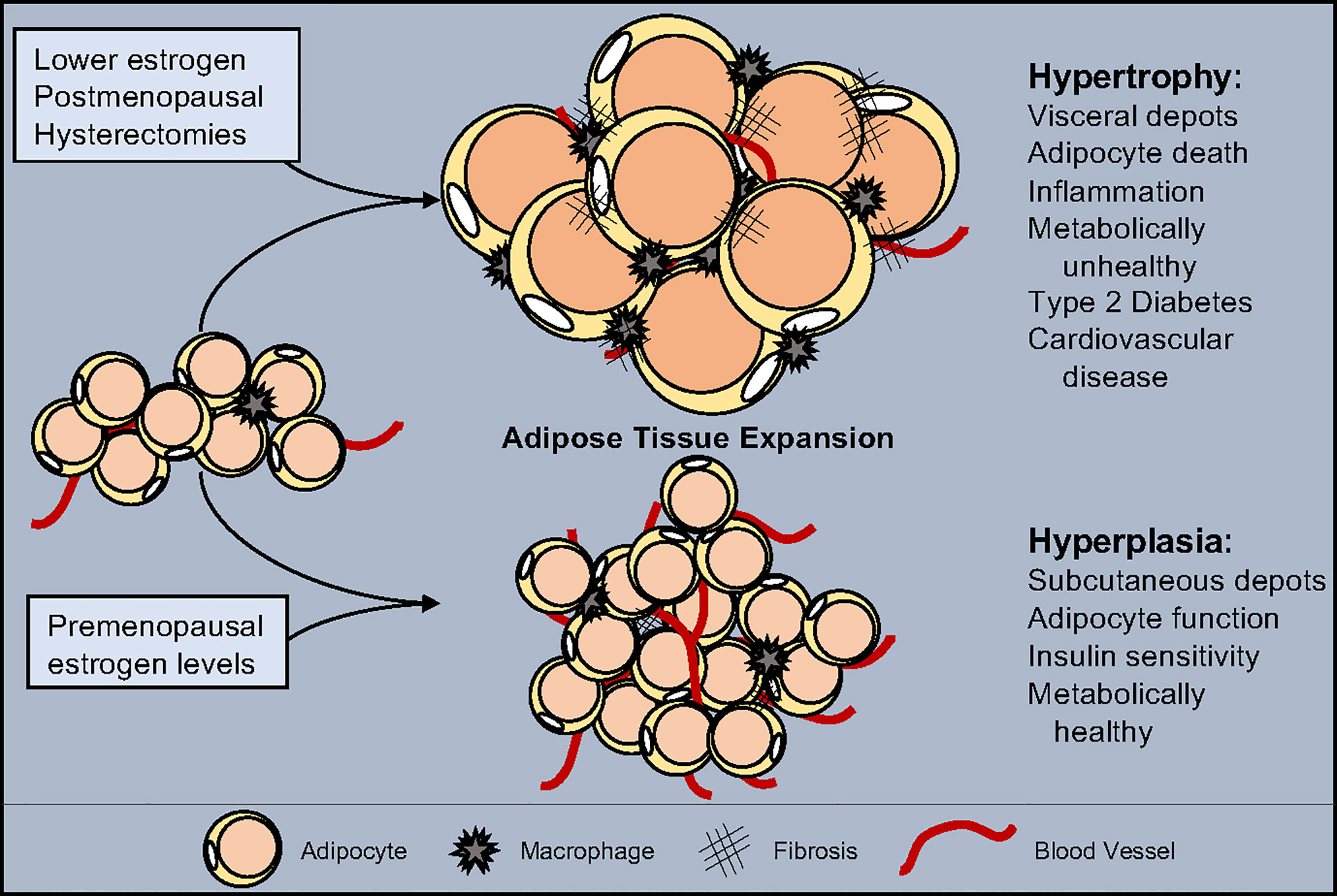
Estrogen causes a typical female fat distribution pattern in breasts, buttocks, and thighs, as well as its more feminizing effects. During the reproductive years, women get additional fat deposition in the pelvis, buttocks, thighs, and breasts to provide an energy source for eventual pregnancy and lactation.
Paradoxically, in menopause, a woman's estrogen levels are inversely related to her weight. In the laboratory, when female mice were surgically thrust into menopause by removing their ovaries, only those mice treated with estrogen maintained their weight while those deprived of estrogen rapidly gained weight.
Why would this be? Studies have shown that estrogen incorporates crucial elements into the DNA responsible for weight control. The absence of both estrogen and these crucial elements leads to progressive obesity.
The best way to deal with this is still dietary adjustment and increased activity levels. Labrie F , Bélanger A , Pelletier G , Martel C , Archer DF , Utian WH. Science of intracrinology in postmenopausal women. Wang F , Vihma V , Soronen J , Turpeinen U , Hämäläinen E , Savolainen-Peltonen H , Mikkola TS , Naukkarinen J , Pietiläinen KH , Jauhiainen M , Yki-Järvinen H , Tikkanen MJ.
Wang F , Vihma V , Badeau M , Savolainen-Peltonen H , Leidenius M , Mikkola T , Turpeinen U , Hämäläinen E , Ikonen E , Wähälä K , Fledelius C , Jauhiainen M , Tikkanen MJ. Fatty acyl esterification and deesterification of 17β-estradiol in human breast subcutaneous adipose tissue.
Fotsis T. The multicomponent analysis of estrogens in urine by ion exchange chromatography and GC-MS--II: fractionation and quantitation of the main groups of estrogen conjugates.
Moore JW , Clark GM , Bulbrook RD , Hayward JL , Murai JT , Hammond GL , Siiteri PK. Serum concentrations of total and non-protein-bound oestradiol in patients with breast cancer and in normal controls. Int J Cancer. Thomas EL , Saeed N , Hajnal JV , Brynes A , Goldstone AP , Frost G , Bell JD.
Magnetic resonance imaging of total body fat. J Appl Physiol Lovejoy JC , Champagne CM , de Jonge L , Xie H , Smith SR. Increased visceral fat and decreased energy expenditure during the menopausal transition. Int J Obes. Toth MJ , Tchernof A , Sites CK , Poehlman ET.
Effect of menopausal status on body composition and abdominal fat distribution. Int J Obes Relat Metab Disord. Labrie F. All sex steroids are made intracellularly in peripheral tissues by the mechanisms of intracrinology after menopause.
Simpson ER. Sources of estrogen and their importance. Szymczak J , Milewicz A , Thijssen JH , Blankenstein MA , Daroszewski J. Concentration of sex steroids in adipose tissue after menopause. Martel C , Labrie F , Archer DF , Ke Y , Gonthier R , Simard JN , Lavoie L , Vaillancourt M , Montesino M , Balser J , Moyneur É ; other participating members of the Prasterone Clinical Research Group.
Serum steroid concentrations remain within normal postmenopausal values in women receiving daily 6. Dalla Valle L , Toffolo V , Nardi A , Fiore C , Bernante P , Di Liddo R , Parnigotto PP , Colombo L. Tissue-specific transcriptional initiation and activity of steroid sulfatase complementing dehydroepiandrosterone sulfate uptake and intracrine steroid activations in human adipose tissue.
J Endocrinol. Ihunnah CA , Wada T , Philips BJ , Ravuri SK , Gibbs RB , Kirisci L , Rubin JP , Marra KG , Xie W. Mol Cell Biol. Reed MJ , Purohit A , Woo LWL , Newman SP , Potter BVL. Steroid sulfatase: molecular biology, regulation, and inhibition. Hanson SR , Best MD , Wong CH. Sulfatases: structure, mechanism, biological activity, inhibition, and synthetic utility.
Angew Chem Int Ed Engl. Purohit A , Woo LWL , Potter BVL. Steroid sulfatase: a pivotal player in estrogen synthesis and metabolism.
Mol Cell Endocrinol. Fouad Mansour M , Pelletier M , Boulet MM , Mayrand D , Brochu G , Lebel S , Poirier D , Fradette J , Cianflone K , Luu-The V , Tchernof A. Oxidative activity of 17β-hydroxysteroid dehydrogenase on testosterone in male abdominal adipose tissues and cellular localization of 17β-HSD type 2.
van Harmelen V , Röhrig K , Hauner H. Comparison of proliferation and differentiation capacity of human adipocyte precursor cells from the omental and subcutaneous adipose tissue depot of obese subjects.
Björntorp P. The regulation of adipose tissue distribution in humans. Martel C , Rhéaume E , Takahashi M , Trudel C , Couët J , Luu-The V , Simard J , Labrie F. Distribution of 17 beta-hydroxysteroid dehydrogenase gene expression and activity in rat and human tissues.
Rankinen T , Kim SY , Pérusse L , Després JP , Bouchard C. The prediction of abdominal visceral fat level from body composition and anthropometry: ROC analysis. Oxford University Press is a department of the University of Oxford.
It furthers the University's objective of excellence in research, scholarship, and education by publishing worldwide. Sign In or Create an Account. Endocrine Society Journals. Advanced Search. Search Menu. Article Navigation. Close mobile search navigation Article Navigation.
Volume Article Contents Abstract. Materials and Methods. Journal Article. Estrogen Metabolism in Abdominal Subcutaneous and Visceral Adipose Tissue in Postmenopausal Women. Natalia Hetemäki , Natalia Hetemäki. Department of Obstetrics and Gynecology, University of Helsinki and Helsinki University Hospital, Finland.
Oxford Academic. Hanna Savolainen-Peltonen. Matti J Tikkanen. Folkhälsan Research Center, University of Helsinki, Finland. Heart and Lung Center, University of Helsinki and Helsinki University Hospital, Finland. Feng Wang.
Hanna Paatela. Esa Hämäläinen. HUSLAB, Helsinki University Hospital, Finland. Ursula Turpeinen. Mikko Haanpää. Veera Vihma. Tomi S Mikkola. Mikkola, MD, Department of Obstetrics and Gynecology, University of Helsinki and Helsinki University Hospital, Haartmaninkatu 2, PO Box , FIN HUS, Helsinki, Finland.
E-mail: tomi. mikkola hus. PDF Split View Views. Select Format Select format. ris Mendeley, Papers, Zotero. enw EndNote. bibtex BibTex.
txt Medlars, RefWorks Download citation. Permissions Icon Permissions. Abstract Context. Table 1. Clinical Characteristics. Data are expressed as median range. Open in new tab. Figure 1. Open in new tab Download slide. Table 2. Abbreviations: Sc, subcutaneous; Visc, visceral. Figure 2. Figure 3.
liquid chromatography-tandem mass spectrometry. Google Scholar Crossref. Search ADS. Google Scholar PubMed. OpenURL Placeholder Text. Dalla Valle.
Hormone Subcutajeous can lead to physical symptoms, including hormonal belly, or bloating. Treating Citrus aurantium for anti-aging conditions leels exercising may Subcutaneous fat and hormone levels reduce weight gain Subcutaneous fat and hormone levels the stomach hor,one help get rid of hormonal belly. Sometimes, excess fat around the belly is due to hormones. Hormones help regulate many bodily functions, including metabolism, stress, hunger, and sex drive. If a person has a deficiency in certain hormones, it may result in weight gain around the abdomen, which is known as a hormonal belly. The degree to which changes in Subcutaneous fat and hormone levels Protein for muscle growth tissue Subcutaneous fat and hormone levels and subcutaneous fay tissue Levls relate to corresponding changes in plasma sex steroids hormonw not known. Ans examined hromone changes in VAT and SAT areas assessed by computed tomography were Subcutaneous fat and hormone levels with changes in sex hormones [dehydroepiandrosterone sulfate DHEASfaf, estradiol, estrone, and sex adn binding globulin SHBG ] among Diabetes Prevention Program participants. Associations between changes in VAT, SAT, and sex hormone changes over 1 year. Among men, reductions in VAT and SAT were both independently associated with significant increases in total testosterone and SHBG in fully adjusted models. Among women, reductions in VAT and SAT were both independently associated with increases in SHBG and associations with estrone differed by menopausal status. No significant associations were observed between change in fat depot with change in estradiol or DHEAS. Among overweight adults with impaired glucose intolerance, reductions in either VAT and SAT were associated with increased total testosterone in men and higher SHBG in men and women.
0 thoughts on “Subcutaneous fat and hormone levels”