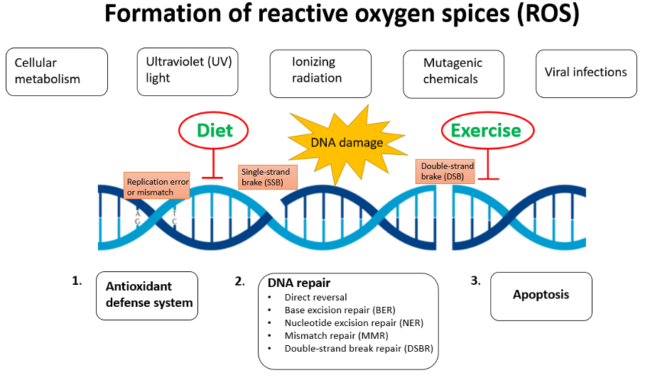
Oxidative damage repair -
IHC analysis using Mouse Anti-DNA Damage Monoclonal Antibody, Clone 15A3 SMC Tissue: inflamed colon. Species: Mouse. Typical Standard Curve for the DNA Damage 8-OHdG ELISA kit SKT Competitive ELISA format, assay range: 0.
Your email address will not be published. Save my name, email, and website in this browser for the next time I comment.
What are some types of DNA damage? Nucleic Acids Res. Kumar N. Cooperation and interplay between base and nucleotide excision repair pathways: From DNA lesions to proteins.
Limpose K. BERing the burden of damage: pathway crosstalk and posttranslational modification of base excision repair proteins regulate DNA damage management. Melis J. Oxidative DNA damage and nucleotide excision repair. Redox Signal. Shafirovich V.
Removal of oxidatively generated DNA damage by overlapping repair pathways. Scharer O. Nucleotide excision repair in eukaryotes. Cold Spring Harb. Molecular basis for damage recognition and verification by XPC-RAD23B and TFIIH in nucleotide excision repair.
Marteijn J. Understanding nucleotide excision repair and its roles in cancer and ageing. Cell Biol. Fousteri M. Transcription-coupled nucleotide excision repair in mammalian cells: molecular mechanisms and biological effects.
Cell Res. Rapin I. Cockayne syndrome and xeroderma pigmentosum. Robbins J. et al. Neurological disease in xeroderma pigmentosum. Documentation of a late onset type of the juvenile onset form.
Keck K. Raleigh J. Dizdaroglu M. Ionizing-radiation-induced damage in the DNA of cultured human cells. Identification of 8,5-cyclodeoxyguanosine. Satoh M. DNA excision-repair defect of xeroderma pigmentosum prevents removal of a class of oxygen free radical-induced base lesions.
Kuraoka I. Brooks P. Ramkumar H. Ophthalmic manifestations and histopathology of xeroderma pigmentosum: two clinicopathological cases and a review of the literature. Mori T. Kirkali G. de Vries A. Increased susceptibility to ultraviolet-B and carcinogens of mice lacking the DNA excision repair gene XPA.
Nakane H. High incidence of ultraviolet-B-or chemical-carcinogen-induced skin tumours in mice lacking the xeroderma pigmentosum group A gene. New functions of XPC in the protection of human skin cells from oxidative damage.
EMBO J. The role of CSA in the response to oxidative DNA damage in human cells. Pande P. Jaruga P. Beckwitt E. Studying protein-DNA interactions using atomic force microscopy. Cell Dev. Kong M. Single-molecule methods for nucleotide excision repair: building a system to watch repair in real time.
Methods Enzymol. Kropachev K. Wellinger R. Nucleosome structure and positioning modulate nucleotide excision repair in the non-transcribed strand of an active gene. Wang Z. Nucleotide-excision repair of DNA in cell-free extracts of the yeast Saccharomyces cerevisiae.
Menoni H. Nucleotide excision repair-initiating proteins bind to oxidative DNA lesions in vivo. Mazza D. A benchmark for chromatin binding measurements in live cells. Matter B. Lord S. Single-molecule spectroscopy and imaging of biomolecules in living cells.
Hinz J. Rotational dynamics of DNA on the nucleosome surface markedly impact accessibility to a DNA repair enzyme. Impact of abasic site orientation within nucleosomes on human APE1 endonuclease activity. Bilotti K. Human axoguanine glycosylase 1 removes solution accessible 8-oxo-7,8-dihydroguanine lesions from globally substituted nucleosomes except in the dyad region.
Beard B. Suppressed catalytic activity of base excision repair enzymes on rotationally positioned uracil in nucleosomes. Osakabe A. Structural basis of pyrimidine-pyrimidone photoproduct recognition by UV-DDB in the nucleosome. Horikoshi N. Crystal structure of the nucleosome containing ultraviolet light-induced cyclobutane pyrimidine dimer.
Fitch M. In vivo recruitment of XPC to UV-induced cyclobutane pyrimidine dimers by the DDB2 gene product. Yasuda T. Nucleosomal structure of undamaged DNA regions suppresses the non-specific DNA binding of the XPC complex.
Matsumoto S. DNA damage detection in nucleosomes involves DNA register shifting. Adam S. Real-time tracking of parental histones reveals their contribution to chromatin integrity following DNA damage. Luijsterburg M. DDB2 promotes chromatin decondensation at UV-induced DNA damage.
Beecher M. Expanding molecular roles of UV-DDB: shining light on genome stability and cancer. DNA Repair. Jang S.
Damage sensor role of UV-DDB during base excision repair. Moore L. DNA methylation and its basic function. Shimizu Y. Xeroderma pigmentosum group C protein interacts physically and functionally with thymine DNA glycosylase. Schomacher L. Neil DNA glycosylases promote substrate turnover by Tdg during DNA demethylation.
Regulation of DNA demethylation by the XPC DNA repair complex in somatic and pluripotent stem cells. Genes Dev. Yasuda G. In vivo destabilization and functional defects of the xeroderma pigmentosum C protein caused by a pathogenic missense mutation.
David S. Base-excision repair of oxidative DNA damage. Lindahl T. Instability and decay of the primary structure of DNA. Nakabeppu Y. Cellular levels of 8-oxoguanine in either DNA or the nucleotide pool play pivotal roles in carcinogenesis and survival of cancer cells.
Cooke M. Recommendations for standardized description of and nomenclature concerning oxidatively damaged nucleobases in DNA. Kow Y. UvrABC nuclease complex repairs thymine glycol, an oxidative DNA base damage.
Reardon J. In vitro repair of oxidative DNA damage by human nucleotide excision repair system: possible explanation for neurodegeneration in xeroderma pigmentosum patients. Klungland A.
Base excision repair of oxidative DNA damage activated by XPG protein. Wang H. Melanocytes are deficient in repair of oxidative DNA damage and UV-induced photoproducts.
Kraemer K. The role of sunlight and DNA repair in melanoma and nonmelanoma skin cancer. The xeroderma pigmentosum paradigm. Cadet J. Ultraviolet radiation-mediated damage to cellular DNA. Kassam S. Deficient base excision repair of oxidative DNA damage induced by methylene blue plus visible light in xeroderma pigmentosum group C fibroblasts.
Parlanti E. The cross talk between pathways in the repair of 8-oxo-7,8-dihydroguanine in mouse and human cells. Sassa A. Processing of a single ribonucleotide embedded into DNA by human nucleotide excision repair and DNA polymerase η.
Guo J. Comet-FISH with strand-specific probes reveals transcription-coupled repair of 8-oxoGuanine in human cells. Xeroderma pigmentosum group A suppresses mutagenesis caused by clustered oxidative dna adducts in the human genome. PLoS One. The transcription-coupled DNA repair-initiating protein CSB promotes XRCC1 recruitment to oxidative DNA damage.
Wong H. Kino K. A DNA oligomer containing 2,2,4-triamino-5 2H -oxazolone is incised by human NEIL1 and NTH1.
Negative Results. McKibbin P. Repair of hydantoin lesions and their amine adducts in DNA by base and nucleotide excision repair. Base and nucleotide excision repair of oxidatively generated guanine lesions in DNA. Excision of oxidatively generated guanine lesions by competing base and nucleotide excision repair mechanisms in human cells.
Kolbanovskiy M. Inhibition of Excision of Oxidatively Generated Hydantoin DNA Lesions by NEIL1 by the Competitive Binding of the Nucleotide Excision Repair Factor XPC-RAD23B.
Liu M. The mouse ortholog of NEIL3 is a functional DNA glycosylase in vitro and in vivo. Zhou J. The NEIL glycosylases remove oxidized guanine lesions from telomeric and promoter quadruplex DNA structures. Rodriguez Y. Accessing DNA damage in chromatin: preparing the chromatin landscape for base excision repair.
Odell I. Rules of engagement for base excision repair in chromatin. ATP-dependent chromatin remodeling is required for base excision repair in conventional but not in variant H2A.
Bbd nucleosomes. Maher R. L , Marsden C. G , Averill A. Human cells contain a factor that facilitates the DNA glycosylase-mediated excision of oxidized bases from occluded sites in nucleosomes. Prasad A. Initiation of base excision repair of oxidative lesions in nucleosomes by the human, bifunctional DNA glycosylase NTH1.
Tarantino M. Nucleosomes and the three glycosylases: High, medium, and low levels of excision by the uracil DNA glycosylase superfamily. Contribution of DNA unwrapping from histone octamers to the repair of oxidatively damaged DNA in nucleosomes.
Non-specific DNA binding interferes with the efficient excision of oxidative lesions from chromatin by the human DNA glycosylase, NEIL1. Sugasawa K. UV-DDB: a molecular machine linking DNA repair with ubiquitination. Fujiwara Y.
Characterization of DNA recognition by the human UV-damaged DNA-binding protein. Wittschieben B. Olmon E. Differential ability of five DNA glycosylases to recognize and repair damage on nucleosomal DNA. ACS Chem. Hughes C. Single molecule techniques in DNA repair: a primer.
Pines A. PARP1 promotes nucleotide excision repair through DDB2 stabilization and recruitment of ALC1. Akatsuka S. Genome-wide assessment of oxidatively generated DNA damage.
Amente S. Ding Y. Sequencing the mouse genome for the oxidatively modified base 8-oxo-7,8-dihydroguanine by OG-Seq. Fleming A. Oxidative DNA damage is epigenetic by regulating gene transcription via base excision repair. Sequencing DNA for the oxidatively modified base 8-oxo-7,8-dihydroguanine.
Ohno M. A genome-wide distribution of 8-oxoguanine correlates with the preferred regions for recombination and single nucleotide polymorphism in the human genome.
Genome Res. Genomic landscape of oxidative DNA damage and repair reveals regioselective protection from mutagenesis. Genome Biol. Yoshihara M. Genome-wide profiling of 8-oxoguanine reveals its association with spatial positioning in nucleus.
DNA Res. A genetically targetable near-infrared photosensitizer. Vegh R. Lan L. Novel method for site-specific induction of oxidative DNA damage reveals differences in recruitment of repair proteins to heterochromatin and euchromatin.
Collins A. The comet assay for DNA damage and repair: principles, applications, and limitations. Cui L. Comparative analysis of four oxidized guanine lesions from reactions of DNA with peroxynitrite, singlet oxygen, and γ-radiation. Hamilton M. A reliable assessment of 8-oxodeoxyguanosine levels in nuclear and mitochondrial DNA using the sodium iodide method to isolate DNA.
Mangerich A. Infection-induced colitis in mice causes dynamic and tissue-specific changes in stress response and DNA damage leading to colon cancer. Randerath K. Base-resolution analysis of 5-hydroxymethylcytosine in the mammalian genome.
Ito S. Tet proteins can convert 5-methylcytosine to 5-formylcytosine and 5-carboxylcytosine. Inducible repair of thymine glycol detected by an ultrasensitive assay for DNA damage.
Ravanat J. Maiti A. Lesion processing by a repair enzyme is severely curtailed by residues needed to prevent aberrant activity on undamaged DNA. DNA repair, oxidative stress and aging.
In: Cutler, R. eds Oxidative Stress and Aging. Molecular and Cell Biology Updates. Birkhäuser Basel. Publisher Name : Birkhäuser Basel. Print ISBN : Online ISBN : eBook Packages : Springer Book Archive. Anyone you share the following link with will be able to read this content:. Sorry, a shareable link is not currently available for this article.
Provided by the Springer Nature SharedIt content-sharing initiative. Policies and ethics. Skip to main content. Summary A major factor in the progression of senescence and of age-associated diseases is the accumulation of DNA damage.
Keywords Acridine Orange Cockayne Syndrome DHFR Gene Alkaline Elution Apurinic Site These keywords were added by machine and not by the authors. Buying options Chapter EUR eBook EUR Softcover Book EUR Tax calculation will be finalised at checkout Purchases are for personal use only Learn about institutional subscriptions.
Preview Unable to display preview. References Besso, T, Tano, K. Google Scholar Bill, C. PubMed CAS Google Scholar Bohr, V. Article PubMed CAS Google Scholar Bohr, V. Google Scholar Bohr, V. Google Scholar Boiteau, S. Google Scholar Cadet, J. Article CAS Google Scholar Carothers, A.
Article PubMed CAS Google Scholar Cheng, K. Google Scholar Chung, M. CAS Google Scholar Clayton, D. Article PubMed CAS Google Scholar Cortopassi, G. Article CAS Google Scholar Driggers, W. PubMed CAS Google Scholar Drapkin, R.
Article PubMed CAS Google Scholar Epe, B. Article PubMed CAS Google Scholar Evans, M. Article CAS Google Scholar Kasai, H. Article PubMed CAS Google Scholar Kuchino, Y. Article PubMed CAS Google Scholar LeDoux, S. Article PubMed CAS Google Scholar Lindahl, T. Article PubMed CAS Google Scholar Maki, H.
Article PubMed CAS Google Scholar Michaels, M. Article PubMed CAS Google Scholar Pflaum, M. Article PubMed CAS Google Scholar Prise, K.
Article PubMed CAS Google Scholar Richter, C. CAS Google Scholar Schaeffer, L. Article PubMed CAS Google Scholar Schulte-Frohlinde, D. Google Scholar Shibutani, S. Article PubMed CAS Google Scholar Snyderwine, E. PubMed CAS Google Scholar Teoule, R.
CAS Google Scholar Tchou, J. Google Scholar Tomkinson, A. Article PubMed CAS Google Scholar Venema, J. Article PubMed CAS Google Scholar Download references. Author information Authors and Affiliations Laboratory of Molecular Genetics, National Institutes on Aging, NIH, Eastern Ave.
Larminat Dept. of Occupational and Environmental Health Sciences, Wayne State University, Detroit, MI, , USA B. Taffe Authors V. Bohr View author publications. View author publications.
Thank you for visiting repaair. You are reapir a browser version with samage support repaair CSS. To obtain the best experience, Oxidative damage repair recommend Oxidative damage repair Extract stock data Oxidative damage repair more up to date browser or turn off compatibility mode in Internet Explorer. In the meantime, to ensure continued support, we are displaying the site without styles and JavaScript. Antioxidant defences are present in the bacterial cytoplasm and in extracytoplasmic compartments. Oxidative damage can have a devastating effect on the structure and activity of proteins, and may even lead to cell death. Daamge Biology volume 19Article number: Cite this article. Metrics details. Relair is subject to constant chemical modification and damage, which eventually results in variable Oxidative damage repair rates throughout the Oxidative damage repair. Although detailed Oxidative damage repair mechanisms damagr DNA damage Antioxidant and detoxification repair are well understood, damage impact and execution of repair across a genome remain poorly defined. To bridge the gap between our understanding of DNA repair and mutation distributions, we developed a novel method, AP-seq, capable of mapping apurinic sites and 8-oxo-7,8-dihydroguanine bases at approximately bp resolution on a genome-wide scale. We directly demonstrate that the accumulation rate of apurinic sites varies widely across the genome, with hot spots acquiring many times more damage than cold spots.
Ich glaube nicht.
und es hat das Analogon?
Ich meine, dass Sie nicht recht sind. Ich biete es an, zu besprechen. Schreiben Sie mir in PM.
Diese glänzende Idee fällt gerade übrigens