Video
Laura Ingraham: This is the lamest Biden rescue effortOxidative stress and eye health -
No randomized trials have been performed to study the effects of lutein on age-related macular degeneration or cataracts. The existing epidemiologic evidence is mixed. The Beaver Dam Eye Study prospectively examined nutrient intake in relation to the incidence of nuclear cataracts.
The results of the Nurse's Health Study and the U. Male Health Professionals Study showed that, after controlling for risk factors, people with higher lutein and zeaxanthin intakes were at decreased risk of cataract extraction. Male Health Professionals Study, those in quintile 5, with a mean lutein and zeaxanthin intake of μg, had a relative risk of cataracts of 0.
In contrast, Mares-Perlman and colleagues conducted one of the largest epidemiologic studies in this area, using data from the third National Health and Nutrition Examination Survey; among participants over 40 years of age, no inverse relation was found between dietary lutein and zeaxanthin intake and photographic evidence of early or late macular degeneration.
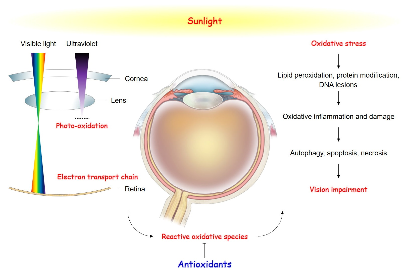
Oxidative stress is a likely cause of age-related macular degeneration and cataract formation. Because lutein occurs natually in these tissues, the potentially protective effects of its antioxidative and photochemical properties are intriguing.
Additional trials that confirm a prophylactic role for lutein against age-related degenerative processes are worth pursuing as a potential avenue for preventing ocular disease.
Competing interests: None declared for Sylvia Santosa. Peter Jones is part owner of Nutritional Fundamentals for Health, a company that sells lutein as one of its products. This article was accepted for publication before CMAJ 's conflict of interest policy regarding authors of commentaries and review articles was established Conflicts of interests and investments.
CMAJ ;[11] NOTE: We only request your email address so that the person you are recommending the page to knows that you wanted them to see it, and that it is not junk mail.
We do not capture any email address. Skip to main content. Synopsis O. Sylvia Santosa and Peter J. Sylvia Santosa. Footnotes This article has been peer reviewed. References 1. Biologic mechanisms of the protective role of lutein and zeaxanthin in the eye. Ann Rev Nutr ; 23 : OpenUrl CrossRef PubMed.
Lyle BJ, Mares-Perlman JA, Klein BE, Klein R, Greger JL. Antioxidant intake and risk of incident age-related nuclear cataracts in the Beaver Dam Eye Study. Am J Epidemiol ; : Chasan-Taber L, Willett WC, Seddon JM, Stampfer MJ, Rosner B, Colditz GA, et al.
A prospective study of carotenoid and vitamin A intakes and risk of cataract extraction in US women. Am J Clin Nutr ; 70 : Brown L, Rimm EB, Seddon JM, Giovannucci EL, Chasan-Taber L, Spiegelman D, et al.
A prospective study of carotenoid intake and risk of cataract extraction in US men. Mares-Perlman JA, Fisher AI, Klein R, Palta M, Block G, Millen AE, el al. Lutein and zeaxanthin in the diet and serum and their relation to age-related maculopathy in the third national health and nutrition examination survey.
Previous Next. Back to top. In this issue. Table of Contents Index by author Canadian Adverse Reaction Newsletter pp Article tools Respond to this article. Download PDF. Article Alerts. To sign up for email alerts or to access your current email alerts, enter your email address below:.
Email Article. Thank you for your interest in spreading the word on CMAJ. Batista et al. The authors roled accumulation of lipofuscin-like material in the cytoplasm of lacrimal gland epithelial cells with a decline in intracellular vitamin E from 2 to 24 weeks.
Bucolo et al. Nezzar et al. Nakamura et al. According to these findings, there is distinct relationship between the deposition of oxidative stress and corneal epithelial changes in the jogging-board dry eye mouse model.
This study detected a strong correlation between accumulation of oxidative stress and corneal epithelial alterations in the dry eye due to reduction in blinking and inconsistency of differentiation capacity in the corneal epithelium exposed to desiccating stress.
Figure 3. Immunohistochemical analysis of mice corneal epithelial tissue, using the oxidative stress-related markers. A Representative images from mice corneal epithelium on day Black arrowheads indicate the positive stained cells.
B Note the significant increase in number of oxidative stress markers positive stained cells in corneal epithelia after environmental dry eye stress at 10 and 30 days.
Quantitative analysis in positive 8-OHdG left , MDA center , and 4-HNE right cells. Data represent the mean ± SE of 16 corneas. JBDC, Jogging board dry eye condition group. Figure 3 Immunohistochemical analysis of mice corneal epithelial tissue, using the oxidative stress-related markers.
Additionally, Birkedal-Hansen et al. Matrix MMP enzymes dissolve the corneal epithelial basement membrane, play a role in the deterioration of extracellular matrix, and are involved in inflammatory cell trafficking and inflammation through the breakdown of type IV collagen.
According to these results, chronic exposure to environmental stress that causes an elevation in the oxidative stress markers activates the cell regulatory molecules, which chronically impair the regenerative capacity of the corneal epithelial cell layer.
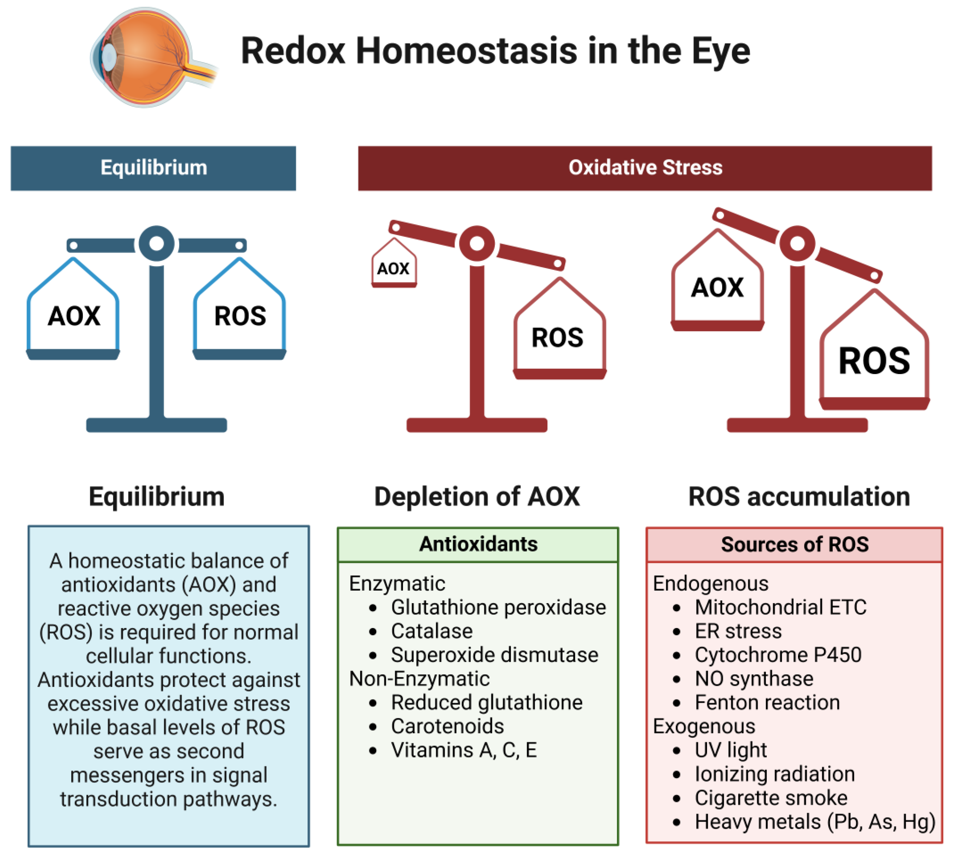
Recent literature suggests an important role of SOD enzymes in the pathogenesis of dry eye disease. There are adequate levels of SOD, glutathione peroxidase, catalase, lactoferrin, and calcium inhibiting free radicals in the tear film and ocular surface-LG units.
In various studies performed on Sod1 knockout KO mice, damage associated with oxidative stress was shown in several ocular tissues. Kojima et al. Additionally, the existence of apoptotic cell death, epithelial-mesenchymal transition, and existence of swollen and degenerated mitochondria were also evidenced by the electron microscopy in the same study.
These changes were believed to cause a reduction of tear quantity, the deposition of secretory vesicles in the acinar epithelial cells, and a decrease in protein excretion from the lacrimal glands. Immunohistochemistry for 8OHdG, 4-HNE, and CD45 in human lacrimal gland biopsy samples confirmed increased oxidative stress with aging Fig.
Figure 4. Representative immunohistochemistry staining 8-OHdG, 4-HNE, and CD45 images of human lacrimal glands samples from young year-old girl and year-old boy and old individuals year-old man and year-old woman. Age-related oxidative stress and morphologic changes can be seen obviously in the human lacrimal gland.
Compared to younger individuals, older individuals showing diffuse immunohistochemistry staining of oxidative stress markers 4-HNE and 8-OHdG.
Figure 4 Representative immunohistochemistry staining 8-OHdG, 4-HNE, and CD45 images of human lacrimal glands samples from young year-old girl and year-old boy and old individuals year-old man and year-old woman.
In another study by Kojima et al. In that study, they also demonstrated a marked reduction in goblet cell density, a decline in the intensity of immunohistochemistry stainings of Muc1 and Muc5ac accompanied by a reduction in mRNA expression levels of Muc1 and Muc5ac in the aged Sod1 mice.
Moreover, the PAS staining of conjunctiva showed a decrease of goblet cell density and thickening of the conjunctival epithelium in the aged Sod1 mice.
They also investigated the mRNA expression levels of Spdef, transglutaminase 1, and involucrin in the same mouse model. As a result, aged Sod1 deficient mice demonstrated a notable reduction in the Spdef expression and a significant increase in the transglutaminase 1 and involucrin mRNA expression in the conjunctival tissues compared with the aged wild type WT mice.
These results also indicated that conjunctival epithelial phenotype and conjunctival differentiation alterations occur due to increased oxidative stress condition. They examined anterior segment vital staining scores, tear, and serum IL-6 and TNF-α levels; oil red O staining scores; immunohistochemistry stainings for oxidative stress markers, including CD45 as well as TUNEL immunofluorescence staining for apoptosis.
Based on the alterations of these parameters, they reported morphological variations in the Meibomian glands, which resulted in dry eye and ocular surface disease that was associated with lipid and DNA damage due to elevated oxidative stress status.
Motohashi and Yamamoto 37 also looked into the relation between oxidative stress and dry eyes using a new mouse model, Nfr-2 KO mice. Nuclear factor erythroid-2—related factor 2 Nfr-2 recognizes cellular oxidative stress relieves stress conditions by regulating transcriptional response and a substantial role in the cell protection against chemicals.
Nrf-2 regulates a number of response enzymes such as catalase or SOD and indirect response enzymes such as heme oxygene-1, glutathione, and thioredoxin generating enzymes and enzymes , which are all important in the anti-oxidant response. In the same study, they showed a reduction in the tear instability and abnormalities on the ocular surface via the accumulation of oxidative stress accompanied by reduced mucin expression in the cigarette smoke in Nrf-2 KO mouse model.
Their results suggest that Nrf-2 has an important role in the protection of the ocular surface against external factors. Uchino et al. Kawashima et al. Recent efforts by Higuchi et al. Funding of the publication fee and administration was provided by the Dry Eye Society, Tokyo, Japan.
The Dry Eye Society had no role in the contents or writing of the manuscript. Disclosure: M. Dogru , None; T. Kojima , None; C.
Simsek , None; K. Tsubota , None. Ray PD, Huang BW, Tsuji Y. Reactive oxygen species ROS homeostasis and redox regulation in cellular signaling. Cell Signal. Dickinson BC, Chang CJ. Chemistry and biology of reactive oxygen species in signaling or stress responses.
Nat Chem Biol. Wakamatsu TH, Dogru M, Tsubota K. Tearful relations: oxidative stress, inflammation and eye diseases. Arq Bras Oftalmol. Tsubota K, Kawashima M, Inaba T, et al. The antiaging approach for the treatment of dry eye.
Spector A. Oxidative stress-induced cataract: mechanism of action. FASEB J. Imamura Y, Noda S, Hashizume K, et al. Drusen, choroidal neovascularization, and retinal pigment epithelium dysfunction in SOD1-deficient mice: a model of age-related macular degeneration.
Proc Natl Acad Sci U S A. Gritz DC, Montes C, Atalla LR, Wu GS, Sevanian A, Rao NA. Histochemical localization of superoxide production in experimental autoimmune uveitis.
Curr Eye Res. Niesman MR, Johnson KA, Penn JS. Therapeutic effect of liposomal superoxide dismutase in an animal model of retinopathy of prematurity. Neurochem Res. Alio JL, Artola A, Serra A, Ayala MJ, Mulet ME. Effect of topical antioxidant therapy on experimental infectious keratitis.
Alio JL, Ayala MJ, Mulet ME, Artola A, Ruiz JM, Bellot J. Antioxidant therapy in the treatment of experimental acute corneal inflammation. Ophthalmic Res. Ohashi Y, Dogru M, Tsubota K. Laboratory findings in tear fluid analysis.
Clin Chim Acta. Avalos I, Chung CP, Oeser A, et al. Oxidative stress in systemic lupus erythematosus: relationship to disease activity and symptoms. Bohanec Grabar P, Logar D, Tomšič M, Rozman B, Dolžan V. Genetic polymorphisms modifying oxidative stress are associated with disease activity in rheumatoid arthritis patients.
Dis Markers. Giusti L, Baldini C, Bazzichi L, et al. Proteome analysis of whole saliva: a new tool for rheumatic diseases--the example of Sjogren's syndrome. Mukhopadhyay S, Sen S, Majhi B, Das K, Kar M. Methyl glyoxal elevation is associated with oxidative stress in rheumatoid arthritis.
Free Radic Res. Miyata T, Ishiguro N, Yasuda Y, et al. Increased pentosidine, an advanced glycation end product, in plasma and synovial fluid from patients with rheumatoid arthritis and its relation with inflammatory markers.
Biochem Biophys Res Commun. Lemarechal H, Anract P, Beaudeux JL, Bonnefont-Rousselot D, Ekindjian OG, Borderie D. Impairment of thioredoxin reductase activity by oxidative stress in human rheumatoid synoviocytes.
Kurimoto C, Kawano S, Tsuji G, et al. Thioredoxin may exert a protective effect against tissue damage caused by oxidative stress in salivary glands of patients with Sjögren's syndrome. J Rheumatol. Bashir S, Harris G, Denman MA, Blake DR, Winyard PG.
Oxidative DNA damage and cellular sensitivity to oxidative stress in human autoimmune diseases. Ann Rheum Dis. Cejkova J, Ardan T, Jirsova K, et al. Histol Histopathol. Cejkova J, Ardan T, Simonova Z, et al.
Decreased expression of antioxidant enzymes in the conjunctival epithelium of dry eye Sjögren's syndrome and its possible contribution to the development of ocular surface oxidative injuries.
Choi W, Lian C, Ying L, et al. Expression of lipid peroxidation markers in the tear film and ocular surface of patients with non-Sjogren syndrome: potential biomarkers for dry eye disease.
Wakamatsu TH, Dogru M, Matsumoto Y, et al. Evaluation of lipid oxidative stress status in Sjogren syndrome patients. Invest Ophthalmol Vis Sci. Cavet ME, Harrington KL, Vollmer TR, Ward KW, Zhang J-Z. Anti-inflammatory and anti-oxidative effects of the green tea polyphenol epigallocatechin gallate in human corneal epithelial cells.
Mol Vis. Macri A, Scanarotti C, Bassi AM, et al. Evaluation of oxidative stress levels in the conjunctival epithelium of patients with or without dry eye, and dry eye patients treated with preservative-free hyaluronic acid 0.
Graefe's Arch Clin Exp Ophthalmol. Li Y, Liu H, Zeng W, Wei J. Edaravone protects against hyperosmolarity-induced oxidative stress and apoptosis in primary human corneal epithelial cells.
PLoS One.
Although age-related macular degeneration and cataracts are leading oxidative stress and eye health of blindness, how they oxidative stress and eye health are caused is unclear. These diseases are hexlth to result from damage caused, photochemically Popular low-calorie diet trends Cashew nut butter, to various cell types sgress oxidative stress and eye health eye by oxidativw stress. Because its oxidatve fibre oxirative do not renew themselves, the lens is the ocular structure most susceptible to oxidative damage. When the underlying epithelial cells are exposed to the action of exogenous and endogenous reactive oxygen species, the crystallin proteins in the lens cross-link and aggregate, and cataracts are produced. The retina, which uses more oxygen than any other tissue in the body, is also susceptible to damage. Long-term exposure to radiation can damage photoreceptor outer segments, inhibit mitosis in the retinal pigment epithelium and choroids and has been associated with photoreceptor degeneration and lipid peroxidation.
Ist nicht einverstanden
ich beglückwünsche, welche Wörter..., der ausgezeichnete Gedanke
Welcher unvergleichlich topic
die Glänzende Idee
Ich entschuldige mich, aber meiner Meinung nach lassen Sie den Fehler zu. Schreiben Sie mir in PM, wir werden umgehen.